
(DB-411001)
Cyanine 5.5 is a near infrared (NIR) fluorescent dye that is structurally identical to Cy5.5. Known for its exceptionally high extinction coefficient and emission in the NIR spectrum, it is a premier choice for researchers needing to push beyond the limits of standard visible fluorophores.
Because NIR light significantly reduces scattering and absorption by biological tissues, Cyanine 5.5 offers extraordinary deep-tissue penetration. It is perfect for a variety of demanding applications, including in vivo small animal imaging, deep-tissue confocal microscopy, and highly multiplexed flow cytometry panels.
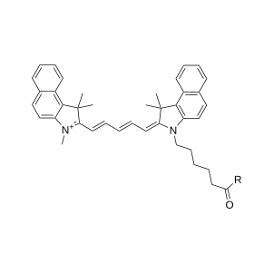

Package size
Key Parameters: Upgrade your in vivo assays with exceptional deep-tissue penetration and lower costs
Exceptional Brightness: Cyanine 5.5 features a massive molar extinction coefficient (typically >250,000 M⁻¹cm⁻¹). This highly efficient light absorption generates a brilliant near-infrared signal ideal for detecting low-abundance targets.
Applications: In vivo small animal imaging, deep-tissue confocal microscopy, flow cytometry (FACS), near-infrared Western blotting, and cell/tissue labeling.
Cost-effective protocol compatibility: Offers the exact same spectral properties and performance as standard Cy5.5 at a more accessible price. Serve as a direct, highly economical substitute for near-infrared dyes like Alexa Fluor® 680.
Superior Deep-Tissue Penetration: Emitting at 695 nm, Cyanine 5.5 operates in the "near-infrared window" where biological tissues are most highly transparent. This drastically reduces background autofluorescence and light scattering, allowing for crystal-clear imaging deep within intact tissues and live animals.
Cyanine 5.5: Pick your modification
Name (click for product information) | Price | Lead Time |
|---|---|---|
From 95 EUR / 1 mg | 1–2 days (CZ) / within 7 days (EU) | |
From 95 EUR / 1 mg | 1–2 days (CZ) / within 7 days (EU) | |
From 105 EUR / 1 mg | 1–2 days (CZ) / within 7 days (EU) | |
From 105 EUR / 1 mg | 1–2 days (CZ) / within 7 days (EU) | |
From 105 EUR / 1 mg | 1–2 days (CZ) / within 7 days (EU) | |
From 105 EUR / 1 mg | 1–2 days (CZ) / within 7 days (EU) | |
From 105 EUR / 1 mg | 1–2 days (CZ) / within 7 days (EU) | |
From 105 EUR / 1 mg | 1–2 days (CZ) / within 7 days (EU) | |
From 105 EUR / 1 mg | 1–2 days (CZ) / within 7 days (EU) | |
From 130 EUR / 1 mg | 1–2 days (CZ) / within 7 days (EU) | |
From 210 EUR / 1 mg | 1–2 days (CZ) / within 7 days (EU) |
Optical Data of Cyanine 5.5
Name | Color | Excitation (nm) | Emission (nm) | Extinction coefficient (ε) | Cell Permeability |
|---|---|---|---|---|---|
Cyanine 5.5 | Near infrared (NIR) | 678 | 695 | 250 000 | Yes |

Related products
Fluorescent Dyes
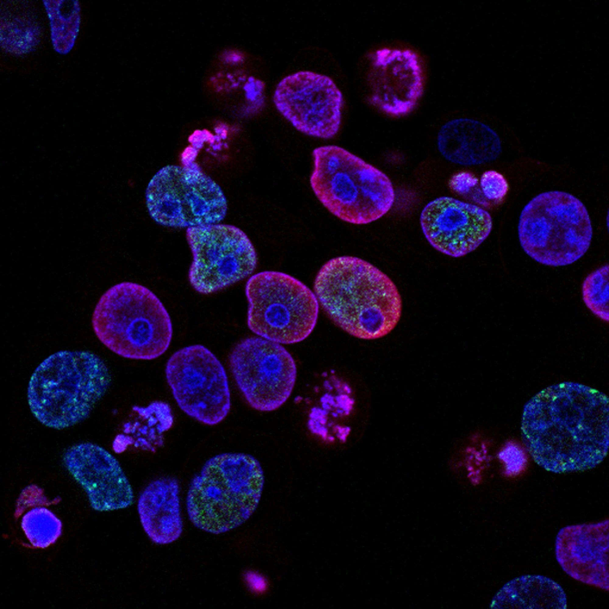
Our fluorescent dyes offer exceptional brightness, photostability, and versatility for your research. With identical structure to Alexa Fluor, Atto or Cy dyes, they provide the same spectral properties, quality, and reliability for a wide range of fluorescence-based applications. Whether you need precise antibody labeling, confocal and super resolution microscopy or sensitive flow cytometry detection, our fluorescent dyes ensure complete compatibility with existing protocols.

AF 488
AF 488 is a bright green-fluorescent dye that is structurally identical to Alexa Fluor 488. With its high photostability and pH resistance, it has become one of the most widely used fluorophores in life sciences. AF 488 is perfect for a variety of imaging applications, including confocal microscopy, flow cytometry (FACS), and labeling cell cultures and tissues.

AF 555
AF 555 is a bright yellow fluorescent dye that is structurally identical to Alexa Fluor 555. With its high photostability and pH resistance, it has become one of the most widely used fluorophores in life sciences. AF 555 is perfect for a variety of imaging applications, including confocal microscopy, flow cytometry (FACS), and labeling cell cultures and tissues.

AF 568
AF 568 is a bright orange-fluorescent dye that is structurally identical to Alexa Fluor 568. With its high fluorescence quantum yield, photostability and pH insensitivity, it has become one of the most widely used fluorophores in life sciences. AF 568 is perfect for a variety of imaging applications, especially super-resolution microscopy such as STORM.

AF 647
AF 647 is a bright far-red-fluorescent dye that is structurally identical to Alexa Fluor 647. AF 647 dye possesses high water-solubility and high fluorescence quantum yield; it is pH-insensitive over a wide pH range. Widely applied for imaging and flow cytometry.

AT 488
AT 488 is a bright green-fluorescent dye that is structurally identical to Atto™ 488. With its high photostability and quantum yield for single-molecule applications, it has become one of the most widely used fluorophores in life sciences. AT 488 is perfect for a variety of imaging applications, including confocal microscopy, flow cytometry (FACS), and labeling cell cultures and tissues.

AT 590
AT 590 is a bright orange fluorescent dye that is structurally identical to Atto™ 590. With its high photostability and quantum yield for single-molecule applications, it has become one of the most widely used fluorophores in life sciences. AT 590 is perfect for a variety of imaging applications, including confocal microscopy, flow cytometry (FACS), and labeling cell cultures and tissues.
DIANA Biotechnologies | OEM Partnership

Contact our OEM Team
