
(DB-411510)
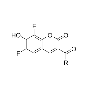
PBlue is a blue fluorescent dye that is structurally identical to Pacific Blue. Its bright fluorescence, high quantum yield, and strong signal at neutral pH make it an excellent choice for flow cytometry, fluorescence microscopy, and biomolecule labeling applications.
Its hydrophilic nature makes it an ideal choice for labeling sensitive proteins. PBlue is perfect for a variety of imaging applications, including confocal microscopy, flow cytometry (FACS), and labeling cell cultures and tissues.
The attached maleimide makes it a thiol-reactive dye for labeling of protein SH groups, and it can be used to attach PBlue fluorophore to proteins and peptides containing cysteine residues, as well as to other thiolated molecules (such as thiol-containing oligonucleotides).

Package size
Key Parameters: Great brightness and pH stability with lower costs
Maleimide: Attaching PBlue to proteins with cystein residues or other thiolated molecules
Applications: Confocal Microscopy, Super Resolution Microscopy, Flow Cytometry (FACS), cell cultures and tissue labeling
Lower Cost Alternative: Same Spectral Properties & Performance – Works exactly like Pacific Blue with friendly price.
Complete Compatibility – Can be used in any existing protocol that uses Pacific Blue
Pricing
Cat. no. | Quantity | Price | Lead Time |
|---|---|---|---|
DB-411510-1MG | 1 mg | 88 EUR | 1–2 days (CZ) / within 7 days (EU) |
DB-411510-5MG | 5 mg | 248 EUR | 1–2 days (CZ) / within 7 days (EU) |
DB-411510-10MG | 10 mg | 371 EUR | 1–2 days (CZ) / within 7 days (EU) |
DB-411510-25MG | 25 mg | 707 EUR | 1–2 days (CZ) / within 7 days (EU) |
DB-411510-50MG | 50 mg | 1194 EUR | 1–2 days (CZ) / within 7 days (EU) |
DB-411510-100MG | 100 mg | 1768 EUR | 1–2 days (CZ) / within 7 days (EU) |
Shipping information
Shipping | Storage | Quality Control |
|---|---|---|
Room Temperature | Up to 24 months in -20 ℃ | HPLC (95 %+ purity) |
Optical Data of PBlue Maleimide
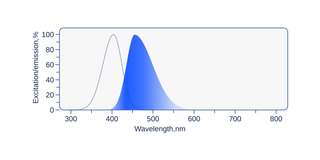
Name | Excitation (nm) | Emission (nm) | Extinction coefficient (ε) |
|---|---|---|---|
PBlue Maleimide | 404 | 455 | 46 000 |
Related products
Fluorescent Dyes
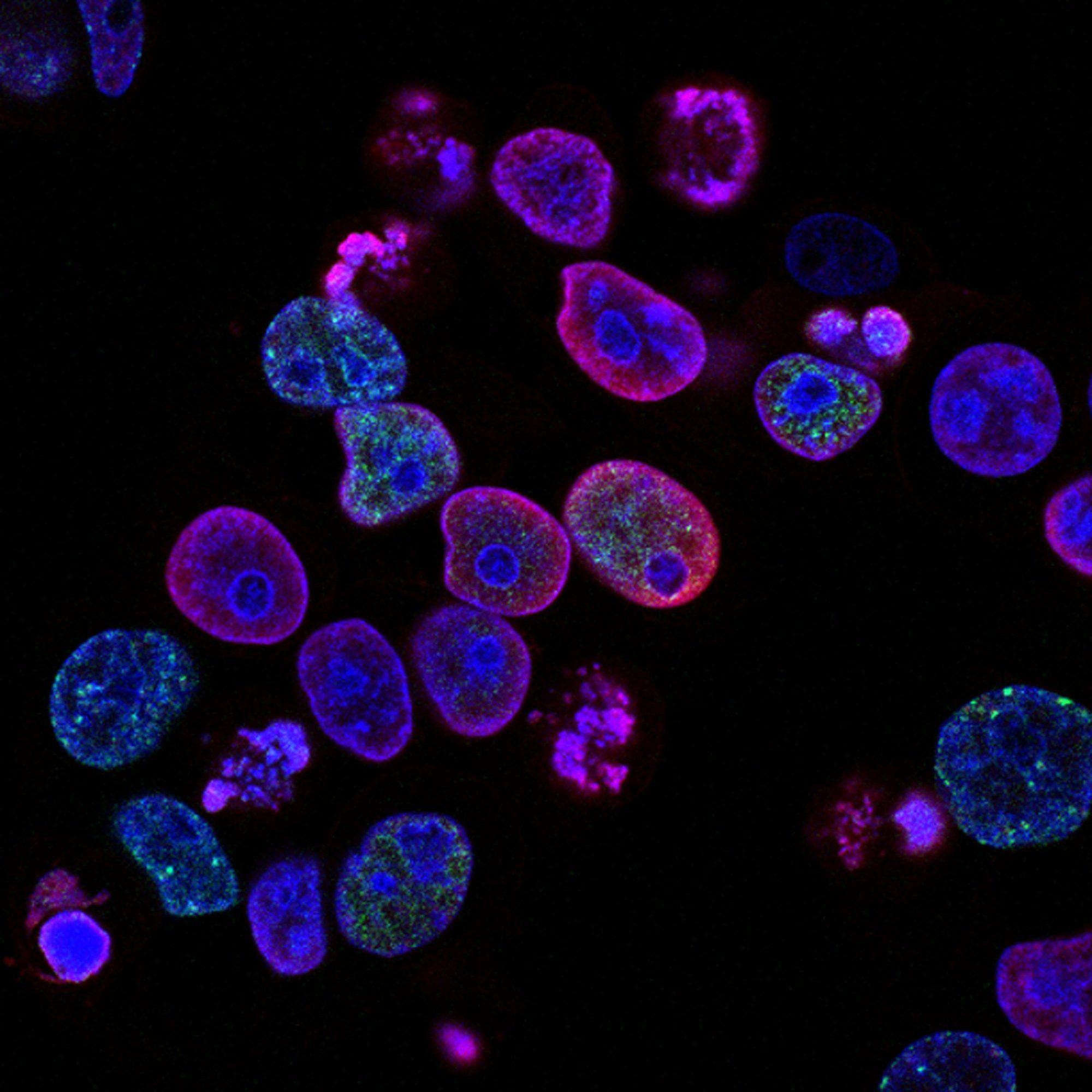
Our fluorescent dyes offer exceptional brightness, photostability, and versatility for your research. With identical structure to Alexa Fluor, Atto or Cy dyes, they provide the same spectral properties, quality, and reliability for a wide range of fluorescence-based applications. Whether you need precise antibody labeling, confocal and super resolution microscopy or sensitive flow cytometry detection, our fluorescent dyes ensure complete compatibility with existing protocols.
PBlue
PBlue is a blue fluorescent dye structurally identical to Pacific Blue™. Its bright fluorescence, high quantum yield, and strong signal at neutral pH make it an excellent choice for flow cytometry, fluorescence microscopy, and biomolecule labeling applications.
AF 700 Maleimide
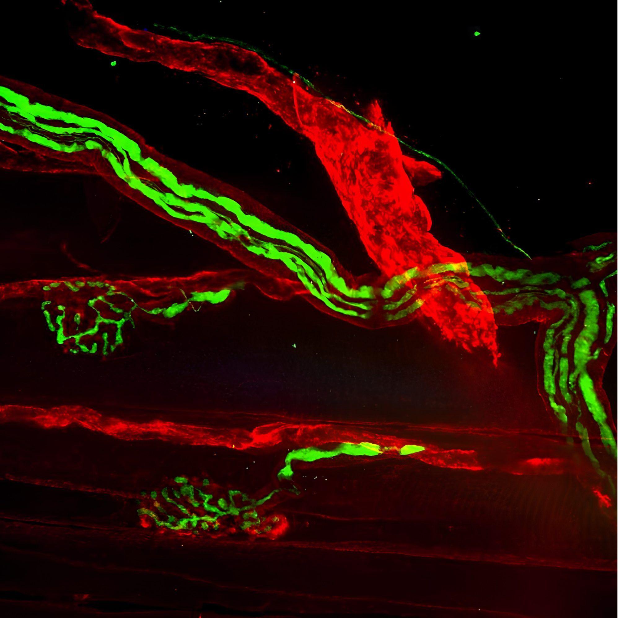
AF 700 Maleimide is near-infrared fluorescent dye that is structurally identical to Alexa Fluor 700. Its excellent photostability and low background fluorescence make it well suited for multicolor flow cytometry and other fluorescence-based applications. AF 700 is excited by standard 633-640 nm red lasers and enables sensitive detection while preserving visible-spectrum channels for multiplex experiments. AF 700 Maleimide is thiol-rective dye for attaching proteins to cystein residues.
txRED Maleimide
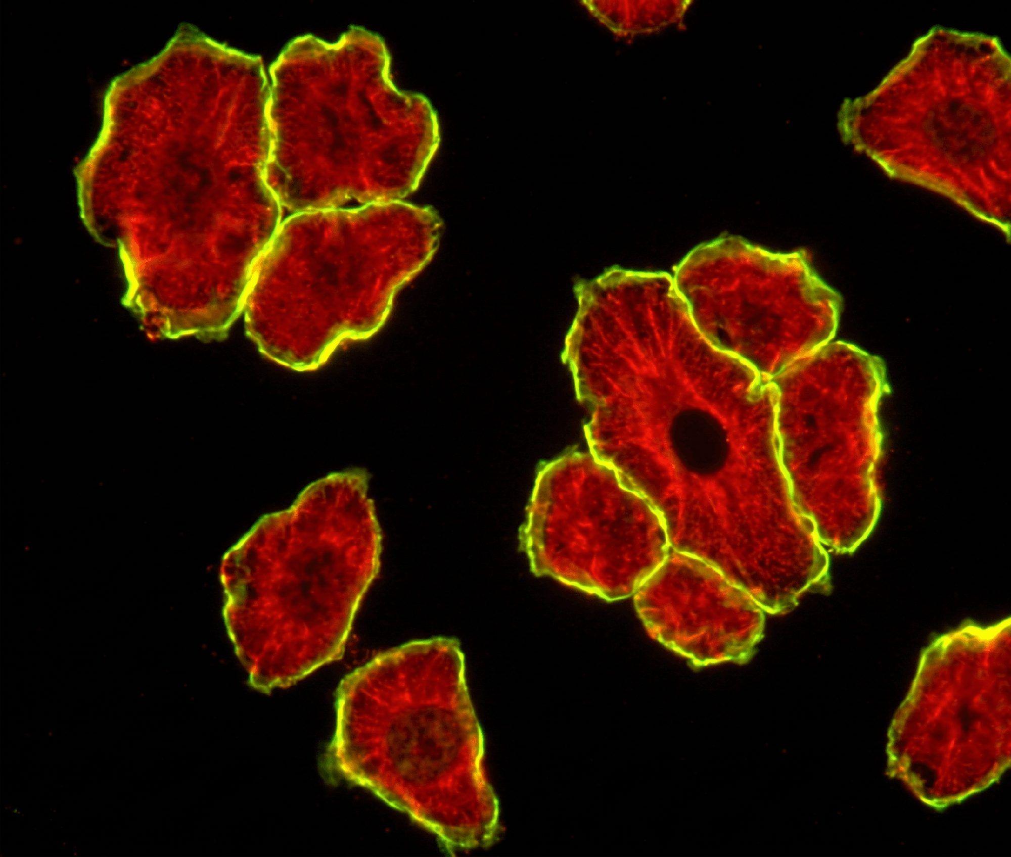
txRED is an orange-red fluorescent dye that is structurally identical to Texas Red-X™. With its high photostability and quantum yield for single-molecule applications, it has become one of the most widely used fluorophores in life sciences. txRED is perfect for a variety of imaging applications, including confocal microscopy, flow cytometry (FACS), and labeling cell cultures and tissues. The attached maleimide makes it a thiol-reactive dye for labeling of protein SH groups, and it can be used to attach AT 488 fluorophore to proteins and peptides containing cysteine residues, as well as to other thiolated molecules (such as thiol-containing oligonucleotides).

Cyanine 5.5 Maleimide
Cyanine 5.5 is a near infrared (NIR) fluorescent dye that is structurally identical to Cy5.5. Known for its exceptionally high extinction coefficient and emission in the NIR spectrum, it is a premier choice for researchers needing to push beyond the limits of standard visible fluorophores. The attached maleimide makes it a thiol-reactive dye for labeling of protein SH groups.

Cyanine 5 Maleimide
Cyanine 5 Maleimide is a far-red fluorescent dye that is structurally identical to Cy5. With its high photostability and quantum yield for single-molecule applications, it has become one of the most widely used fluorophores in life sciences. Cyanine 5 Maleimide is perfect for a variety of imaging applications, including confocal microscopy, flow cytometry (FACS), and labeling cell cultures and tissues. The DBCO allows fluorescent labeling of azide-containing biomolecules inside living cells or whole organisms. The attached maleimide makes it a thiol-reactive dye for labeling of protein SH groups.

AT 590 Maleimide
AT 590 Maleimide is a bright orange-fluorescent dye that is structurally identical to Atto 590 with Maleimide attached. With its high photostability and pH resistance, it has become one of the most widely used fluorophores in life sciences. AT 590 Maleimide is thiol-rective dye for attaching proteins to cystein residues.

AT 488 Maleimide
AT 488 Maleimide is a bright green-fluorescent dye that is structurally identical to Atto 488 with Maleimide attached. With its high photostability and pH resistance, it has become one of the most widely used fluorophores in life sciences. AT 488 Maleimide is thiol-rective dye for attaching proteins to cystein residues.

AF 555 Maleimide
AF 555 Maleimide is a bright yellow-fluorescent dye that is structurally identical to Alexa Fluor 555 with Maleimide attached. With its high photostability and pH resistance, it has become one of the most widely used fluorophores in life sciences. AF 555 Maleimide is thiol-rective dye for attaching proteins to cystein residues.

AF 647 Maleimide
AF 647 Maleimide is a bright far-red-fluorescent dye that is structurally identical to Alexa Fluor 647 Maleimid. With its high photostability and pH resistance, it has become one of the most widely used fluorophores in life sciences. AF 647 Maleimide is thiol-rective dye for attaching proteins to cystein residues.

AF 594 Maleimide
AF 594 Maleimide is a bright red-fluorescent dye that is structurally identical to Alexa Fluor 594. With its high photostability and pH resistance, it has become one of the most widely used fluorophores in life sciences. Its hydrophilic nature makes it an ideal choice for labeling sensitive proteins. AF 594 Maleimide is thiol-rective dye for attaching proteins to cystein residues.

AF 568 Maleimide
AF 568 Maleimide is a bright orange-fluorescent dye that is structurally identical to Alexa Fluor 568. With its high photostability and pH resistance, it has become one of the most widely used fluorophores in life sciences. Its hydrophilic nature makes it an ideal choice for labeling sensitive proteins. AF 568 Maleimide is thiol-rective dye for attaching proteins to cystein residues.
AF 488 Maleimide
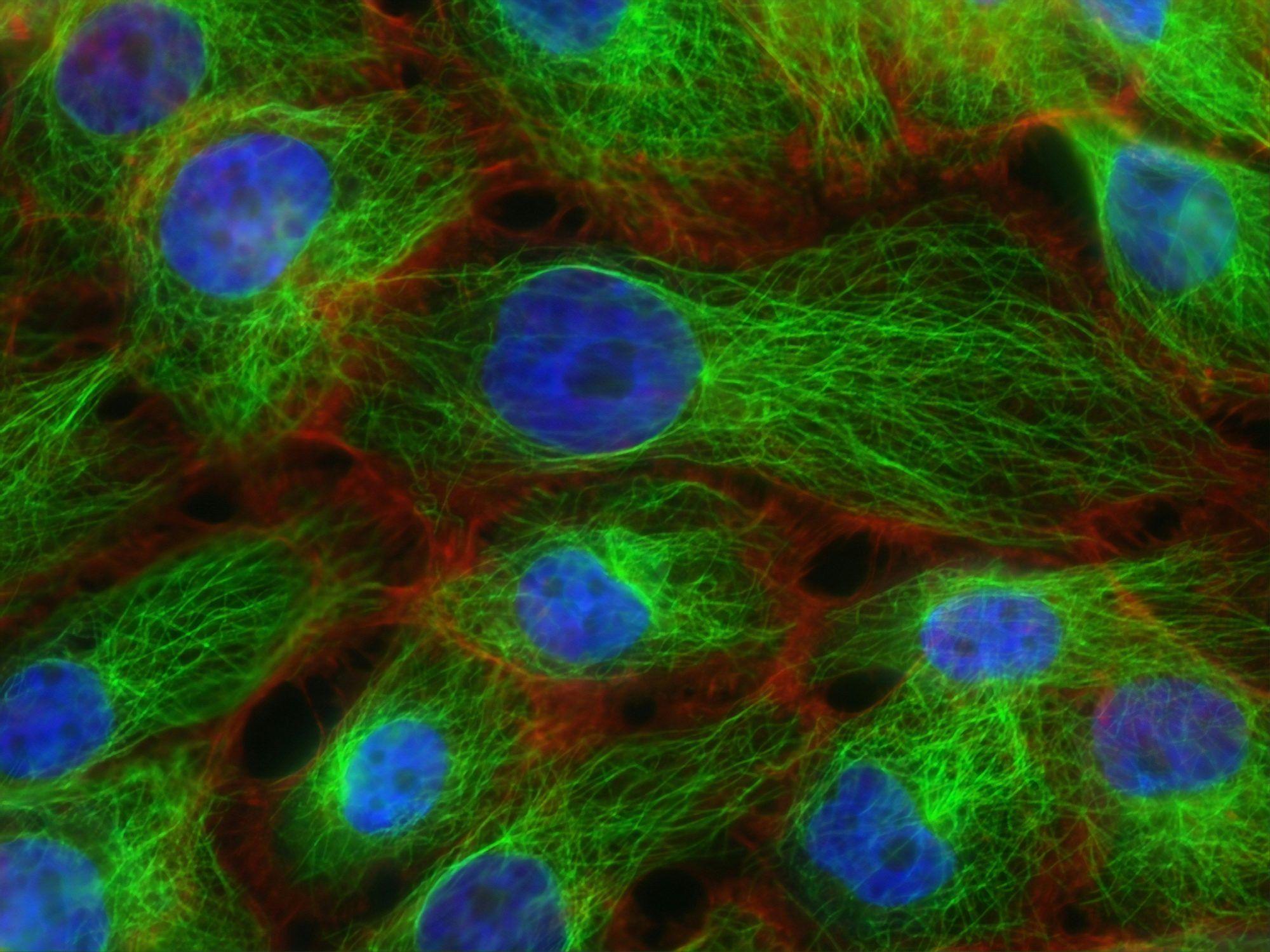
AF 488 Maleimide is a bright green-fluorescent dye that is structurally identical to Alexa Fluor 488 with Maleimide attached. With its high photostability and pH resistance, it has become one of the most widely used fluorophores in life sciences. AF 488 Maleimide is thiol-rective dye for attaching proteins to cystein residues.

AF 350 Maleimide
AF 350 Maleimide is a blue fluorescent dye that is structurally identical to Alexa Fluor 350 with Maleimide attached. With its high photostability and pH resistance, it is widely used in life science research. AF 350 Maleimide is thiol-rective dye for attaching proteins to cystein residues.
DIANA Biotechnologies | OEM Partnership

Contact our OEM Team
