
(DB-411601)
Cyanine 7 is a water-soluble near-infrared (NIR) fluorescent dye that is structurally identical to sulfo-Cy7. With its high extinction coefficient and emission at 773 nm, it reaches far beyond the limits of standard visible fluorophores.
Because NIR light is only weakly absorbed and scattered by biological tissues, Cyanine 7 offers excellent deep-tissue penetration with minimal background autofluorescence. Its sulfonated, hydrophilic nature makes it an ideal choice for labeling sensitive proteins. Cyanine 7 is perfect for a variety of applications, including in vivo small animal imaging, deep-tissue imaging, and near-infrared Western blotting.


Package size
Key Parameters: Upgrade your in vivo assays with deep-tissue NIR imaging and lower costs
Deep-Tissue NIR Imaging: Emitting at 773 nm, Cyanine 7 operates in the near-infrared window where biological tissues are most transparent, drastically reducing autofluorescence and light scattering
Applications: In vivo small animal imaging, deep-tissue imaging, near-infrared Western blotting, and cell/tissue labeling
Lower Cost Alternative: Same Spectral Properties & Performance – Works exactly like sulfo-Cy7 at a more accessible price
Complete Compatibility – Can be used in any existing protocol that uses sulfo-Cy7
Cyanine 7: Pick your modification
Name (click for product information) | Price | Lead Time |
|---|---|---|
Upon Request | 1–2 days (CZ) / within 7 days (EU) |
Optical Data of Cyanine 7
Name | Color | Excitation (nm) | Emission (nm) | Extinction coefficient (ε) | Cell Permeability |
|---|---|---|---|---|---|
Cyanine 7 | Near infrared (NIR) | 750 | 773 | 240 600 | No |

Related products
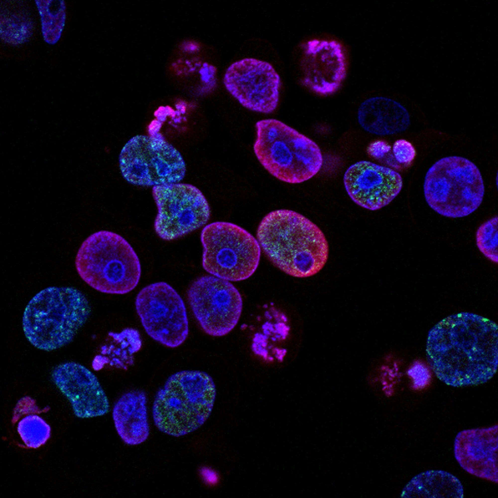
Fluorescent Dyes
Our fluorescent dyes offer exceptional brightness, photostability, and versatility for your research. With identical structure to Alexa Fluor, Atto or Cy dyes, they provide the same spectral properties, quality, and reliability for a wide range of fluorescence-based applications. Whether you need precise antibody labeling, confocal and super resolution microscopy or sensitive flow cytometry detection, our fluorescent dyes ensure complete compatibility with existing protocols.
Cyanine 7 NHS Ester
Cyanine 7 NHS Ester is a water-soluble near-infrared (NIR) fluorescent dye that is structurally identical to sulfo-Cy7 with NHS ester attached. With its high extinction coefficient and emission deep in the near-infrared region, it is a premier choice for imaging where tissue autofluorescence and light scattering must be minimized. Its sulfonated, hydrophilic nature makes it an ideal choice for labeling sensitive proteins in purely aqueous conditions. Cyanine 7 NHS Ester is perfect for a variety of applications, including in vivo small animal imaging, deep-tissue imaging, and near-infrared Western blotting.

Cyanine 5.5
Cyanine 5.5 is a near-infrared (NIR) fluorescent dye that is structurally identical to Cy5.5. With its high photostability and quantum yield for single-molecule applications, it has become one of the most widely used fluorophores in life sciences. Cyanine 5.5 is perfect for a variety of imaging applications, including confocal microscopy, flow cytometry (FACS), and labeling cell cultures and tissues.

Cyanine 5
Cyanine 5 is a far-red fluorescent dye that is structurally identical to Cy5. With its high photostability and quantum yield for single-molecule applications, it has become one of the most widely used fluorophores in life sciences. Cyanine 5 is perfect for a variety of imaging applications, including confocal microscopy, flow cytometry (FACS), and labeling cell cultures and tissues.

AF 647
AF 647 is a bright far-red-fluorescent dye that is structurally identical to Alexa Fluor 647. AF 647 dye possesses high water-solubility and high fluorescence quantum yield; it is pH-insensitive over a wide pH range. Widely applied for imaging and flow cytometry.
DIANA Biotechnologies | OEM Partnership

Contact our OEM Team
